Topo | Atlas 9000 | Tomo | Pentacam | Galilei G4/G6
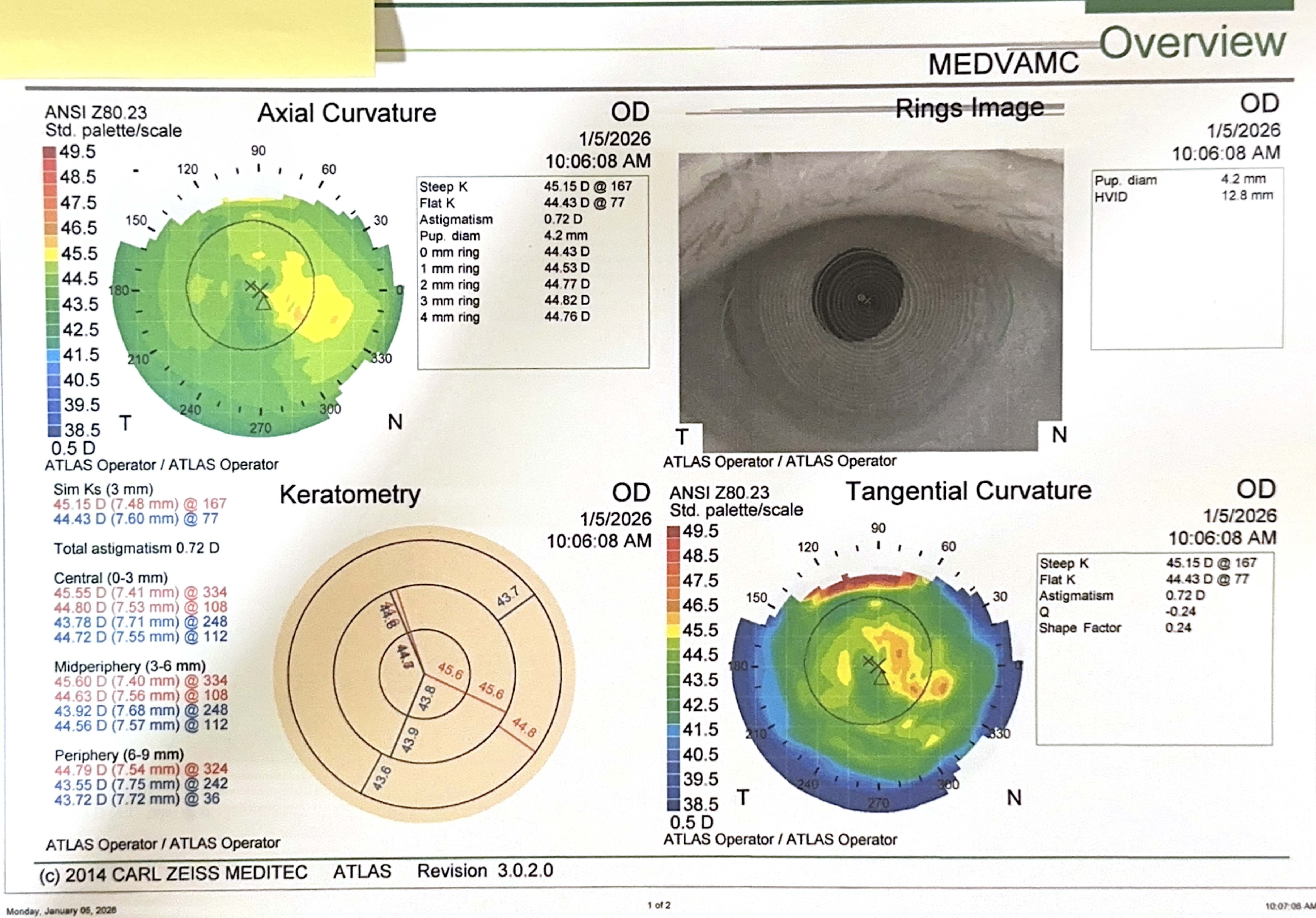
Topography | Atlas 9000
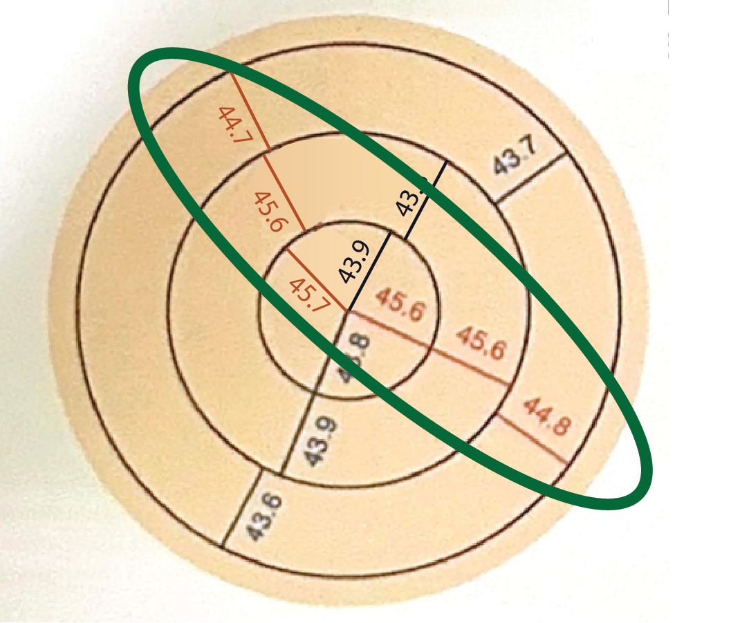
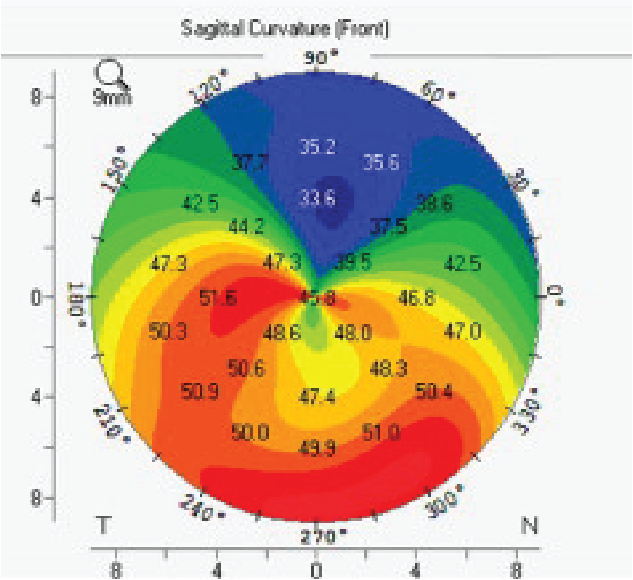
Placido based topographer.
- Maps the anterior corneal surface curvature via keratoscopic rings (mires).
Detects shape of corneal surface, evaluates tear film and corneal surface.
Measures include
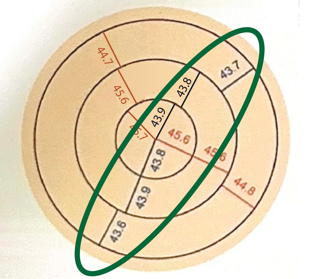
K1: Flat meridian
K2: Steep meridian
Km: Averages of K1 and K2
Astigmatism: Difference between K1 and K2
Q-value: Vector direction and steepness of cornea

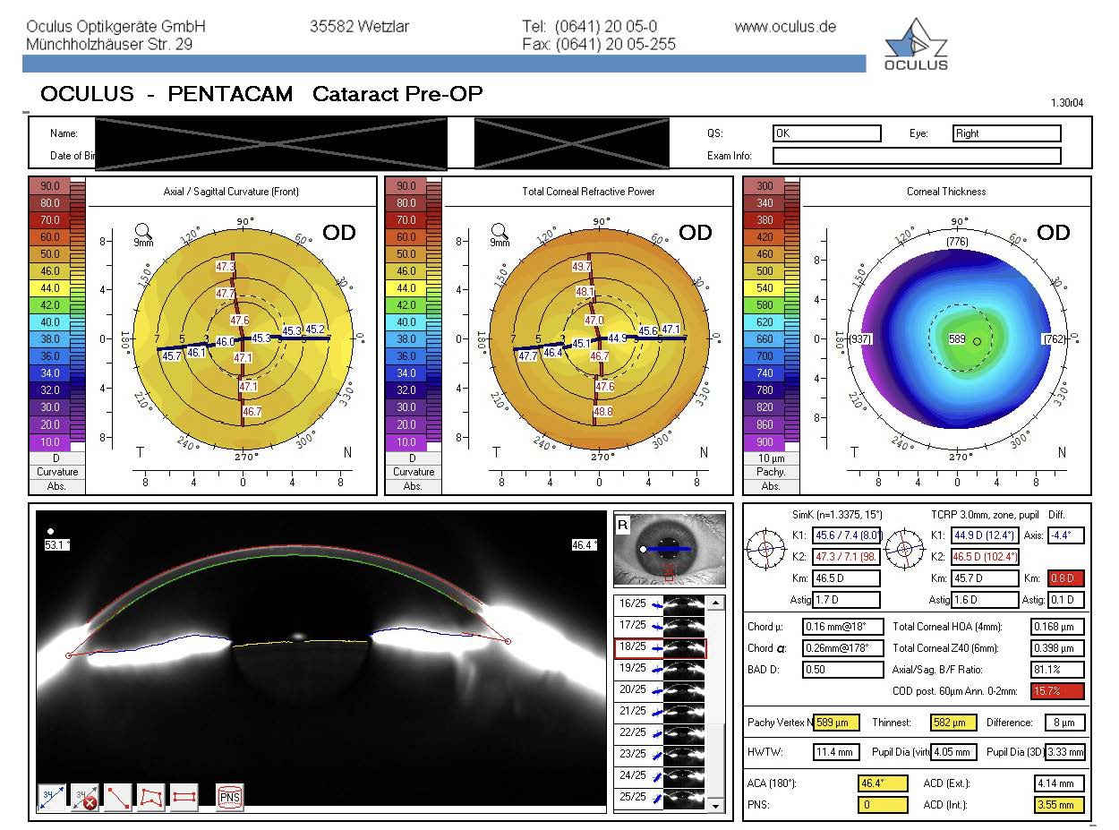
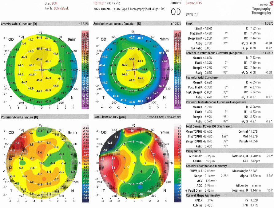
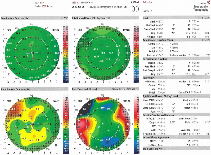
Tomography | Pentacam & Galilei G4/G6
Maps the anterior and posterior corneal surface curvatures.
Maps 3D image of the cornea, enhances ability to detect ectasias.
Pentacam
- Uses Schiempflug based tomographer.
Galilei G4/G6
- Uses a dual Schiempflug based tomographer (has 2 rotating cameras) + Placido based topographer.
Includes all topographic measures and the following
Posterior & Anterior Elevation Data
Corneal Pachymetry across the cornea
Total Corneal Refractive Power
Kmax
Corneal Optic Density score (Pentacam)
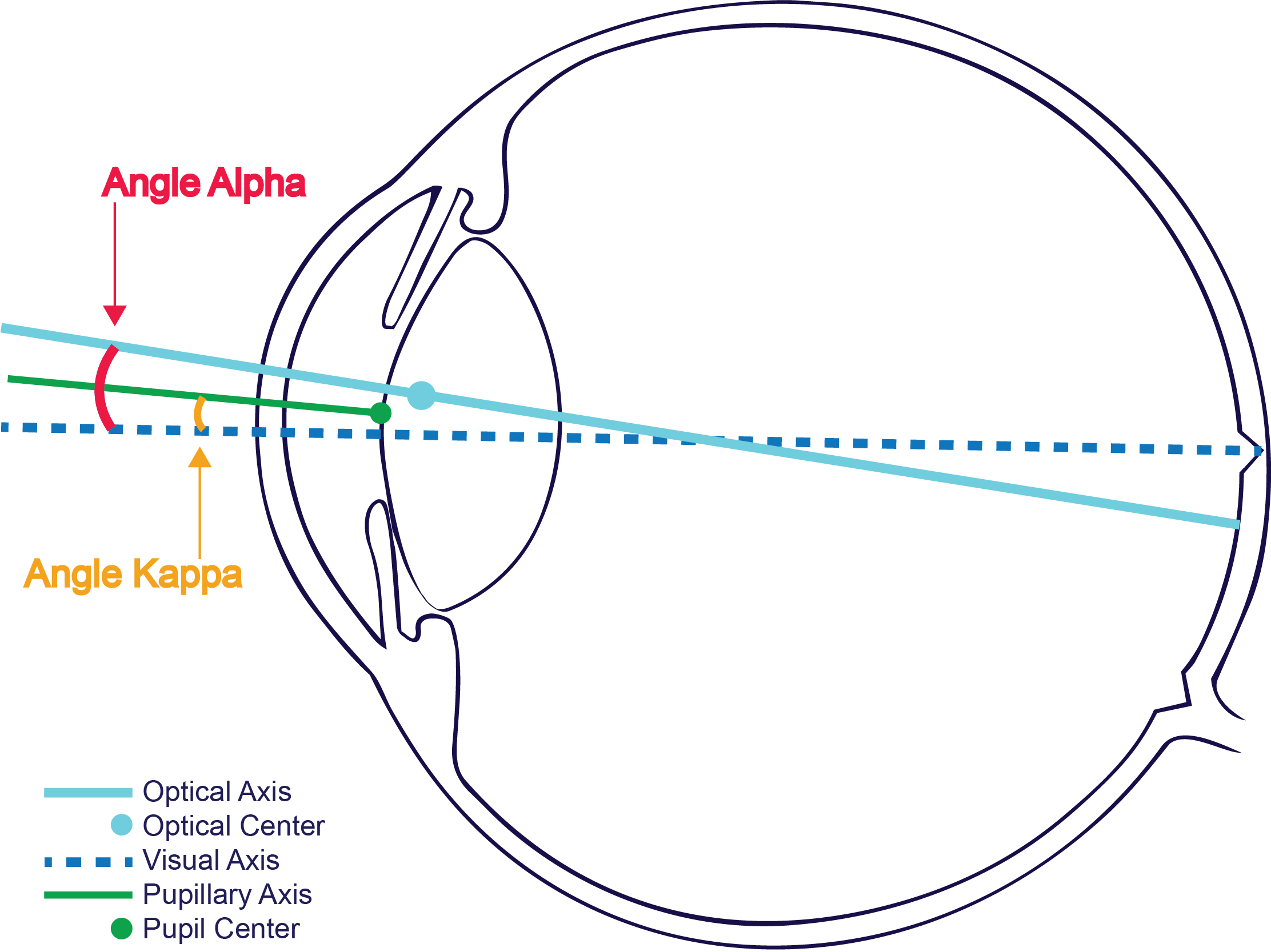
Angle 𝛼
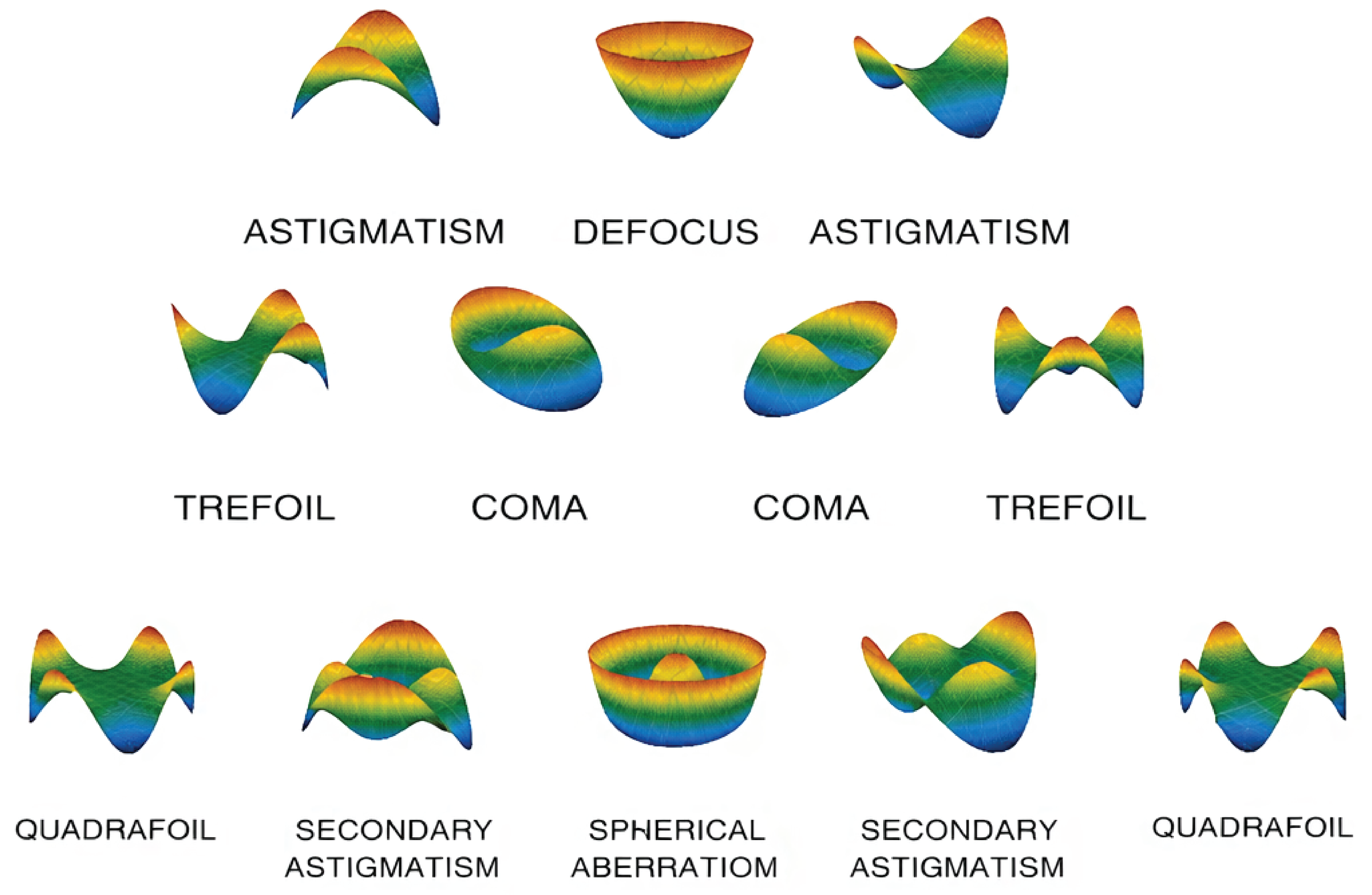
Angle 𝜅Higher Order Aberrations - Including Spherical Aberrations
Pentacam