Measurements for lens selection and intraoperative considerations
Lenstar 900 | IOL Master 700 | ARGOS
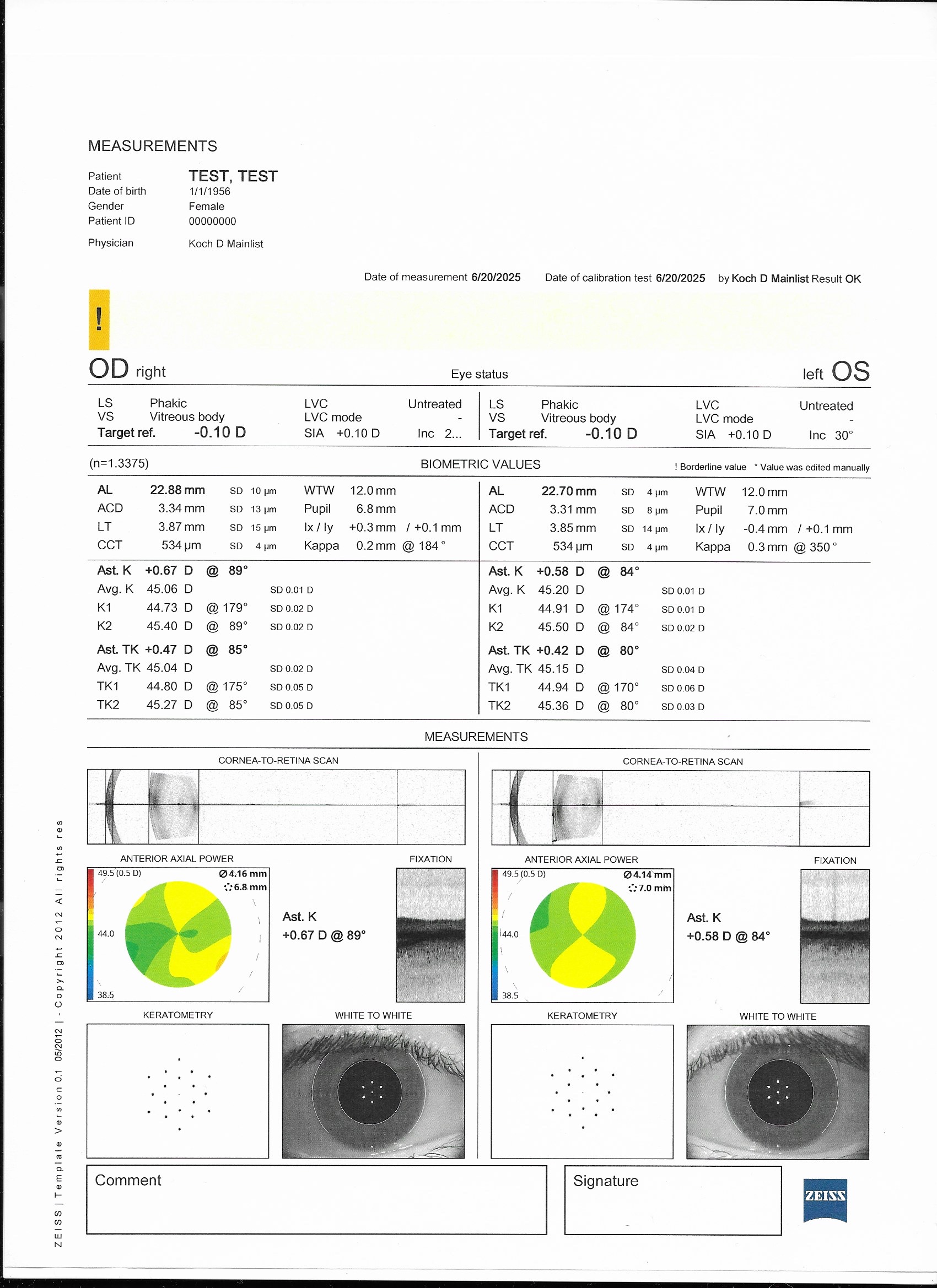
Lens thickness (LT), Central Cornea Thickness (CCT), Axial length (AL), Anterior Chamber Depth (ACD), and Keratometry readings (Ks).
May have inaccurate or inconsistent measurements in eyes with dry eyes, corneal scarring, PSCs, very mature NSCs, or vitreous hemorrhage.
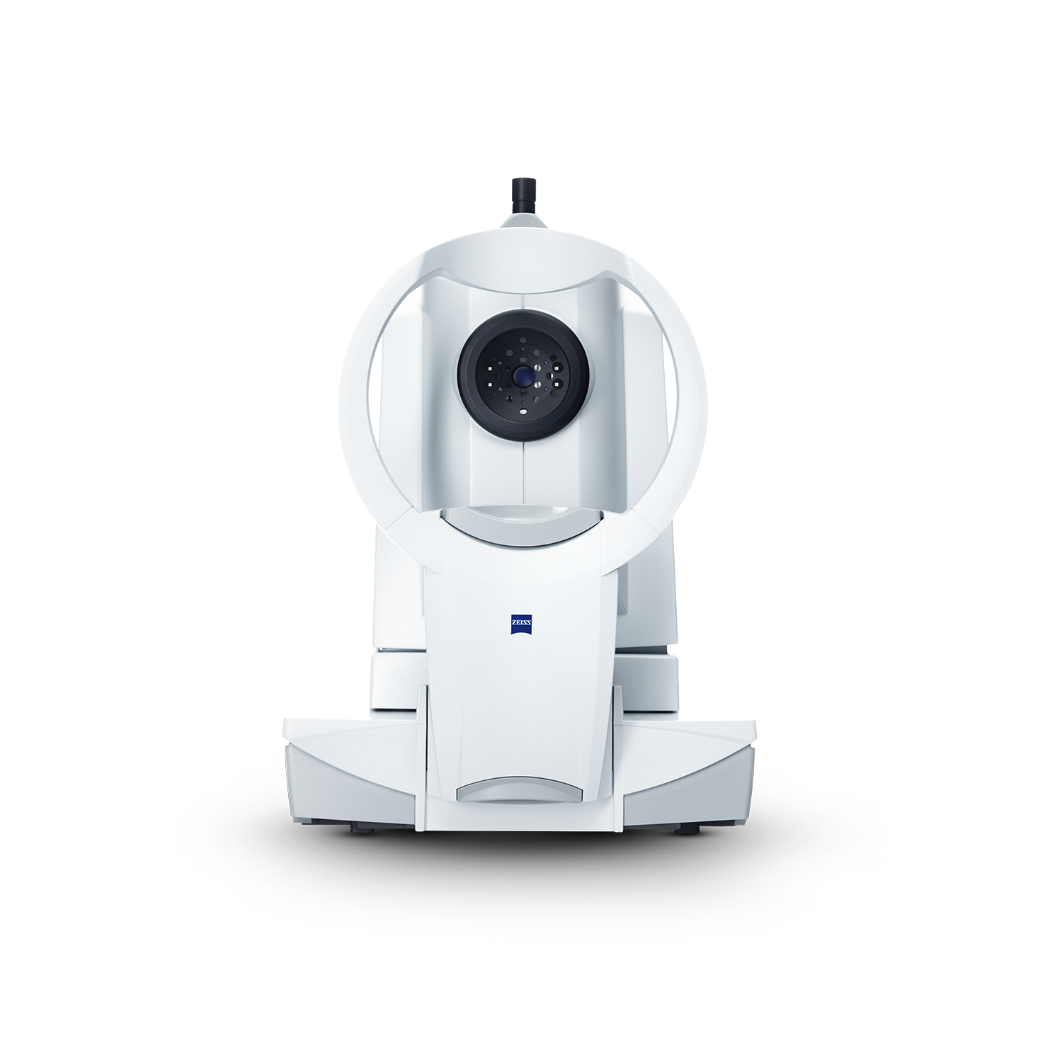
Based on swept source OCT
Better for obtaining measurements in eyes with:
- Dense media (eg, dense cataract)
- Longer axial lengths
Arriola-Villalobos et al 2016
Provides total keratometry (TK) measurements
Direct anterior and posterior corneal curvature.
- Helpful for Toric IOL planning.
**Ensure the IOL formula you put TK into does not already account for posterior corneal astigmatism to avoid overestimating this effect**
Shorter duration to capture scan
Easier for patients with tremors and cognitive impairments to complete.
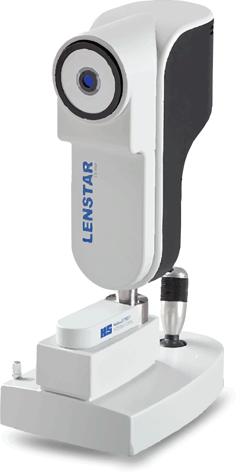
Based on optical low coherence reflectometry
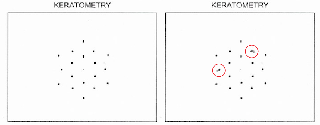
Evaluates 32 points on the cornea.
- Provides more sensitive corneal curvature data.

Based on swept source OCT with segmental refractive index processing
Has an enhanced retinal visualization mode.
- Improves penetration is through dense cataracts.
Anterior corneal surface to macula/point of fixation.
Corneal thickness + Anterior chamber depth + lens thickness + vitreous chamber depth.
Normal: 22.5mm-25mm
Short: <22.5mm
Nanophthalmos: <20mm
- Typically hyperopic patients.
- Always check in eyes that require ≥+2.50 correction.
- This will allow you to plan ahead & avoid complications.
Long: >25.3mm
IOL calculation formulas:
- Barrett Universal
- Holladay I WITH Wang-Koch modification
Prevents IOL power underestimation^
Inter-globe difference ≥ 2mm
Pathologic etiology should be identified or at least ruled out.
Examples:
- Congenital syndromes
- Scleral buckle
- Choroidal mass/thickening
AL off by 1 mm ≈ IOL error of 2.5D
- Overestimated AL → Myopic surprise
- Underestimated AL → Hyperopic surprise
AL Standard Deviation
- Should be <0.02mm
Distance from central corneal epithelium to central corneal endothelium.
Affects accuracy of IOP measurements.
Normal: 520-560µm
Abnormally Thin: <520µm
Risk factors
- Refractive surgery
- KCN & other ectatic disorders
- Connective tissue disorders.
Associated with falsely low IOP readings.
Abnormally Thick: >560µm
Risk factors
- Corneal edema
- Corneal dystrophies
Associated with falsely high IOP readings, poor wound healing, worsening edema.
CCT >640µm
Risk factor for
- Severe postoperative corneal edema, requiring endothelial transplant
Distance from central corneal endothelium to anterior capsule of lens.
Normal: 2.6mm-3.6mm
Abnormally Shallow: <2.6mm
Risk factors
- Zonulopathy
- ACG
- Pupillary block or iris bombe
- Intumescent cataracts
- Anterior inflammation
- Trauma
Abnormally Deep: >3.6mm
Risk factors
- Zonulopathy
- Pigmentary dispersion syndrome
- Plateau iris syndrome
- Trauma
Horizontal diameter of cornea from limbus to limbus.
Normal: 12mm-12.5mm
Used in IOL calculations to predict Effective Lens Position (ELP).
- ELP is the largest variable that cannot be directly measured.
All Biometers provide...
Corneal Power
Average (air-tear-interface): ~42-43D
Anterior surface: ~ (+)48-49D
Air-cornea interface adds convergence.
Posterior surface: ~ (-)6D
Aqueous-cornea interface adds divergence.
Anterior Corneal Surface Measurements
K1: Flat meridian
K2: Steep meridian
Avg K: Average of K1 & K2
- Should be between 40D-47D for each eye
- Difference between eyes should be ≤1D
Astigmatism changes with age; shape of cornea changes with age.
Flat meridian (K1) steepens ~+0.4D every 10 years, while steep meridian (K2) flattens with age.
Keratometry off by 1D ≈ IOL error of 0.9D
- Overestimated K → Hyperopic surprise
- Underestimated K → Myopic surprise
Avg K Standard Deviation
- Should be <0.3D per meridian
IOL Master 700 only...
Total Keratometry (TK)
Accounts for both anterior and posterior corneal surfaces.
Ideal for astigmatic patients interested in toric lenses.
Patients with irregular astigmatism are TYPICALLY NOT toric lens candidates.
See topography/tomography page for more info.
Sources: OKAPP IOL ppt, CBSE Clinical Optics: Corneal Optics, AAO young ophthalmologists: how to read corneal topography
Amount of astigmatism induced by primary incision during cataract surgery.
Calculated by vector analysis
Based on pre- and (surgeon specific) post-op keratometry.
Average SIA: -0.1-0.2 D
Useful for
- Evaluating toric lens necessity
- Predicting amount of post-operative astigmatic correction required.
Factors influencing SIA
1. Incision location
- Primary incision at the cornea induces flattening in the incised meridian and steepening at the meridian 90º away.
Therefore, an incision at the temporal cornea produces mild (+) cylinder at ~180º axis.
An incision at a greater distance from visual axis will have less influnce on a SIA.
2. Incision size
- Larger incision correlates with higher corneal SIA change.
Significantly greater astigmatic changes occur with incision size >3.2mm compared to ≤2.2mm.
3. Incision tunnel length
- Incisions with shorter tunnel lengths have less SIA.
Lenstar 900
Quality is based on the following standard deviations.
AL: <0.02 mm
K1 & K2: <0.3 D
Meridian for astigmatism: <3.5º
IOL Master 700
Quality is based on the following perameters.
Cornea to retina scan
- Can identify presence of lens tilt.
Anterior axial power
- A basic topography to show whether the anterior surface is regular.
Fixation image
- Demonstrates whether patient was fixated correctly when image was taken.
Keratometry image
- Quality can significantly alter reported astigmatism in:
- Magnitude
- Axis
Smearing of just 1 of the 19 reflectance keratometry reflected spots can result in innacurate measurements.
IOL Master 700 Only
Distance between visual axis and optical axis
- Important for evaluating diffractive lens eligibility.
| Ix | + | Iy | ≈ Angle 𝛼
*If the absolute value of Ix or Iy is > 0.3, subtract 0.1 from the sum of above equation.
Angle 𝛼 <0.3
- Can use DO-ATIOLs in these patients.
Angle 𝛼 = 0.3 - 0.5
- Difficult to predict outcome with DO-ATIOLs; caution with these patients.
Angle 𝛼 >0.5
- Avoid DO-ATIOLs.
*Disclaimer: There is still some debate over the true effect of angle alpha on diffractive IOL outcomes but the authors do take this into consideration when planning.
DO-ATIOLs = Diffractive Optic Advanced Technology IOLs.
See Corneal Optics under Key Concepts on Topography/Tomography page.